Back Of Skull And Neck Anatomy - Bones of the Head and Neck | Interactive Anatomy Guide : The large, complex muscles of the neck and back move the head, shoulders, and vertebral column.
byAdmin•
0
Back Of Skull And Neck Anatomy - Bones of the Head and Neck | Interactive Anatomy Guide : The large, complex muscles of the neck and back move the head, shoulders, and vertebral column.. The neck begins at the base of the skull and connects to the thoracic spine (the upper back). Anatomy the base of the skull is a complex area. The majority of these nerves control the functions of the upper extremities and allow you to feel your arms, shoulder, and back of your head. The erector spinae group forms the majority of the muscle mass of the back and it is the primary extensor of the vertebral column. The large, complex muscles of the neck and back move the head, shoulders, and vertebral column.
The skeletal section of the head and neck forms the top part of the axial skeleton and is made up of the skull, hyoid bone, auditory ossicles, and cervical spine. A herniated disc between the vertebrae in your neck can cause extreme pain in the base of your skull and back of your neck if the herniated disc presses on a nerve root. An area called the occiput. Neck muscles are bodies of tissue that produce motion in the neck when stimulated. The neck contains seven of.
Superficial Muscles Posterior View | The Superficial ... from s-media-cache-ak0.pinimg.com Ullrich says that the inflammation in the facet joints in the cervical spine between the shoulders and base of the head causes pain at the back of the head. The neck is connected to the upper back through a series of seven vertebral segments. The majority of these nerves control the functions of the upper extremities and allow you to feel your arms, shoulder, and back of your head. The posterior muscles of the neck are primarily concerned with head movements, like extension. They move the head in every direction, pulling the skull and jaw towards the shoulders, spine, and scapula. Nerves and vessels form neurovascular bundles, which may be harmed at the sites of skull openings by pathological process or trauma. The nerves of the head and neck include the most vital and important organs of the nervous system — the brain and spinal cord — as well as the organs of the special senses. A pinched nerve in your cervical spine is called cervical radiculopathy.
The neck attaches the head to the trunk.
Ullrich says that the inflammation in the facet joints in the cervical spine between the shoulders and base of the head causes pain at the back of the head. It is, therefore, the transitional part of the body between the skull superiorly and the clavicles inferiorly that joins the head to the trunk and limbs. (2) the veins of the neck. The back muscles stabilize and move the vertebral column, and are grouped according to the lengths and direction of the fascicles. The muscles of the neck run from the base of the skull to the upper back and work together to bend the head and. A pinched nerve in your cervical spine is called cervical radiculopathy. An area called the occiput. The neck is connected to the upper back through a series of seven vertebral segments. Muscles of the neck and back. The large, complex muscles of the neck and back move the head, shoulders, and vertebral column. In other words, there is a muscle on the forehead (frontalis) and one on the back of the. The occipital bone is the only bone in your head that connects with your cervical spine (neck). In addition, in this region we also find the major cranial and spinal nerves that connect the central nervous system to the organs, skin, and muscles of the head and neck.
Neck anatomy nerves picture there are 8 spinal nerves that originate from the cervical spine. Anterior fossa, middle fossa, and posterior fossa. The neurocranium (cranial vault) and the viscerocranium (facial skeleton). The neck begins at the base of the skull and connects to the thoracic spine (the upper back). The cervical spine supports the weight and movement of your head and protects the nerves exiting your brain.
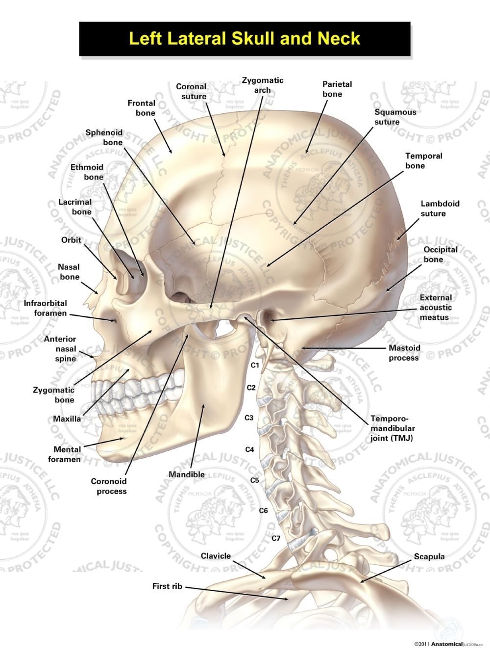
Left Lateral Skull and Neck from anatomicaljustice.com The occipital glands (lymphoglandulæ occipitales), one to three in nu ber, are placed on the back of the head close to the margin of the trapezius and resting on the insertion of the semispinalis capitis.their afferent vessels drain the occipital region of the scalp, while their efferents pass to the superior deep cervical glands. A herniated disc between the vertebrae in your neck can cause extreme pain in the base of your skull and back of your neck if the herniated disc presses on a nerve root. The head rests on the top part of the vertebral column, with the skull joining at c1 (the first cervical vertebra known as the atlas). The veins of the head and neck may be subdivided into three groups: The large, complex muscles of the neck and back move the head, shoulders. The skull can be further subdivided into: It controls flexion, lateral flexion, and rotation of the vertebral column, and maintains the lumbar curve. The posterior auricular glands (lymphoglandulæ auriculares.
The veins of the head and neck may be subdivided into three groups:
Neck muscles are bodies of tissue that produce motion in the neck when stimulated. The neck is the start of the spinal column and spinal cord. The occipital bone is a bone that covers the back of your head; Neck anatomy nerves picture there are 8 spinal nerves that originate from the cervical spine. (2) the veins of the neck. A pinched nerve in your cervical spine is called cervical radiculopathy. It controls flexion, lateral flexion, and rotation of the vertebral column, and maintains the lumbar curve. It involves the upper cervical spine, facet joints, muscles, tendons, ligaments, and nerves. (3) the diploic veins, the veins of the brain, and the venous sinuses of the dura mater.: The large, complex muscles of the neck and back move the head, shoulders. Human skull from the front. They move the head in every direction, pulling the skull and jaw towards the shoulders, spine, and scapula. This example from gray's anatomy shows the cartilages of the throat and the surface anatomy of the neck, with the prominent sternocleidomastoideus which is often thrown into sharp relief when the head is turned or tilted.
It terminates toward the back of the head, behind the ear. The occipital bone is the only bone in your head that connects with your cervical spine (neck). The neck muscles, including the sternocleidomastoid and the trapezius, are responsible for the gross motor movement in the muscular system of the head and neck. In other words, there is a muscle on the forehead (frontalis) and one on the back of the. It controls flexion, lateral flexion, and rotation of the vertebral column, and maintains the lumbar curve.
Dentistry lectures for MFDS/MJDF/NBDE/ORE: Diagrams Of ... from 4.bp.blogspot.com The skull can be further subdivided into: Anatomy of the head and neck. A pinched nerve in your cervical spine is called cervical radiculopathy. The skull base is the inferior portion of the neurocranium. Neck muscles are bodies of tissue that produce motion in the neck when stimulated. Ullrich says that the inflammation in the facet joints in the cervical spine between the shoulders and base of the head causes pain at the back of the head. The neck attaches the head to the trunk. • the external carotid artery is divided into branches (facial, temporal and occipital arteries) which supply the skin and muscles of the face, side and back of the head respectively.
The posterior auricular glands (lymphoglandulæ auriculares.
The back muscles stabilize and move the vertebral column, and are grouped according to the lengths and direction of the fascicles. The neck is connected to the upper back through a series of seven vertebral segments. The neck muscles, including the sternocleidomastoid and the trapezius, are responsible for the gross motor movement in the muscular system of the head and neck. The neurocranium (cranial vault) and the viscerocranium (facial skeleton). The occipital bone is the only bone in your head that connects with your cervical spine (neck). Anatomy the base of the skull is a complex area. The posterior auricular glands (lymphoglandulæ auriculares. These muscles can extend the head, laterally flex it, and rotate it (figure 7). The cavities with the skull muscles in your neck and the top part of your back aren't as large. Back of skull and neck anatomy. The top of the cervical spine connects to the skull, and the bottom connects to the upper back at about shoulder level. • the external carotid artery is divided into branches (facial, temporal and occipital arteries) which supply the skin and muscles of the face, side and back of the head respectively. The occipital glands (lymphoglandulæ occipitales), one to three in nu ber, are placed on the back of the head close to the margin of the trapezius and resting on the insertion of the semispinalis capitis.their afferent vessels drain the occipital region of the scalp, while their efferents pass to the superior deep cervical glands.
The cervical spine supports the weight and movement of your head and protects the nerves exiting your brain back of skull anatomy. Muscles of the neck and back by openstax is licensed under cc by 4.0 ) the erector spinae group forms the majority of the muscle mass of the back and it is the primary extensor of the vertebral column.